
Tesla Stock Is Crashing Real Bad
The global economy is declining today, as companies and investors are trying to empty the so -called “mutual definitions” Donald Trump.
Trump aims to reduce the United States’ dependence on foreign goods by raising the cost of importing for different countries, which aims to the United States’s global supply chain in chaos. Wall Street is in a state of chaos as a result, as the market value has been eliminated about $ 5 trillion in the past two days – the worst performance of the S&P 500 since the epidemic shock for 2020.
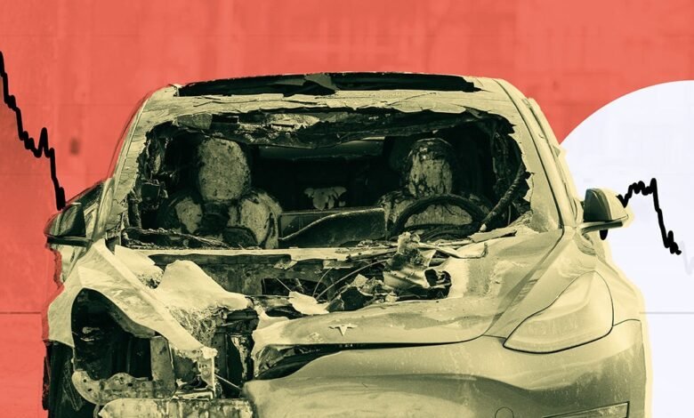
Technology shares were specially serious after China announced its tariffs by 34 percent in response to Trump’s movements, thanks to its dependence on Chinese minerals and work. Among all the technology companies bleeding on the stock exchange, Tesla is especially good.
EV’s EV’s Elon has published losses for ten last 11 week, and this was not different. On Friday, Tesla decreased immediately after China announced its return shot, and continued to decrease throughout the day to a 10 percent decrease. In general, the value of Tesla has decreased by almost 44 percent since Trump took office.
This is before you mention even the results of the quarterly quarterly sales, which came in less than the worst estimates at the lowest level of three years. Analysts now expect a decrease in the second consecutive sales in Tesla, bad news of Tesla’s long -term expectations.
Everything meets together to threaten Tesla as “growth stocks”, a company that wears iron like alphabet or Apple, which is seen as financial growth as guaranteed. Of course, this future depends on the health stock market and confidence, which may not see it again for a while, as all signs indicate stagnation.
Musk’s personal wealth, which is tightly linked to Tasla, has been covered with the same. Thanks to the Trump tariff, his huge reservoir of wealth threw about $ 11 billion, for a loss of 2.5 percent. Forbes I previously mentioned in March that the net value of Musk has decreased by $ 104 billion since Trump took office on January 20, a number that may seem that this customs tariff may seem soon like changing Starlink contracts, and the European Union is its revenge measures.
Since the world is measured for more economic repercussions in the coming weeks, it is clear that Trump’s tariff may have led to a chain reaction that can reshape global markets – and EV’s MUSK – forever.
More about Tesla: The government, which seeks the brutal punishment of the man accused of sabotaging the Tesla Agency
2025-04-05 17:25:00